
Recent studies have shown that CSF flow investigation at the aqueductal and cervical levels could help also to differentiate between these brain disorders, particularly between AD and NPH. Nevertheless, difficulties still exist in the differential diagnosis between atrophy, AD, NPH, or VaD in a nonnegligible part of elderly patients presenting moderate ventricular dilation associated with moderate Hakim triad symptoms. In contrast, one of the common features revealed by MR neuroimaging is the dilatation of the lateral ventricular system, which is more severe in the group with NPH. These three brain disorders (AD, VaD, and NPH) are distinguished by their specific pathological features. During overnight ICP monitoring, many B-waves can be observed, and during infusion study with ICP monitoring, pressure volume compensatory reserve parameters are altered. Nevertheless, it is not a pathology with normal intracranial pressure (ICP) according to Bret et al. NPH is a curable disease associated with ventricular dilation and the presence of a part or total symptoms of Hakim’s triad such as cognitive impairment, gait disturbance, or urinary problems. However, recent population-based studies have estimated the prevalence of NPH to be about 0.5% in those over 65 years old, with an incidence of about 5.5 patients per 100,000 of people per year.

The number of people who develop hydrocephalus or who are currently living with it is difficult to establish since there is no national registry or database of affected people. Studies from various countries found that the proportion of dementia cases diagnosed as vascular dementia ranged from 10 to 38% making it the second most common cause of dementia after Alzheimer’s disease. Its prevalence ranges from 5 to and may vary depending on the studied population. VaD includes all forms of dementia related to cerebrovascular diseases. Recent epidemiological studies predict a doubling of its global prevalence every 20 years, estimated to be actually as high as 24 million affected persons in the world. The most common forms of brain disorders in the elderly regroup Alzheimer’s disease (AD), vascular dementia (VaD), and also normal pressure hydrocephalus (NPH).Īlzheimer disease is the leading cause of dementia and is characterized by a progressive decline in cognitive function, which typically begins with deterioration in memory. It is still not well elucidated if cerebrospinal fluid (CSF) oscillations amplitude can be associated with ventricle dilatations and how its volume variations are implicated in brain disorders. The cerebrospinal fluid fluctuations in brain disorders may depend on many physiological parameters other than the ventricular morphology.

Lateral ventricles volumes are significantly proportional to the 2D reference section area and could be used for patients’ follow-up even if 3D images are unavailable. On the other hand, no significant correlations were detected between ventricles and stroke volumes ( ).

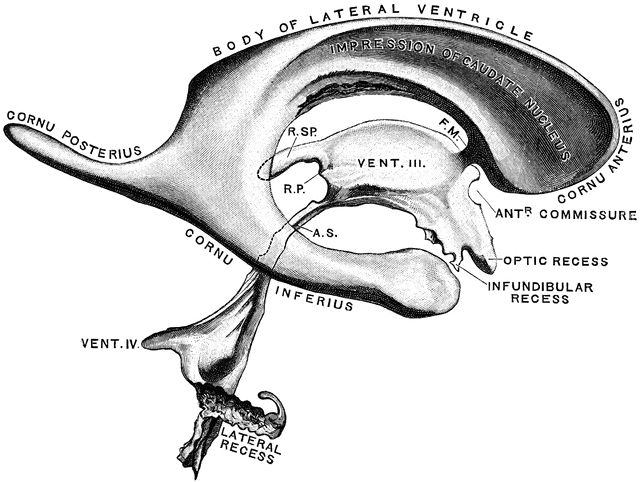
A derived equation was determined to represent the volumes for any given area. The linear regression between volumes and areas was computed, and a strong positive correlation was detected ( ). The largest volumes were observed in hydrocephalus patients. A common reference plane was used to calculate the lateral ventricles’ area on 2D images. Lateral ventricles and stroke volumes were assessed on anatomical and phase contrast scans, respectively. FForty-five elderly patients suffering from Alzheimer’s disease (19), normal pressure hydrocephalus (13), and vascular dementia (13) were involved and underwent anatomical and phase contrast MRI scans. This work suggests a fast estimation method of the lateral ventricles volume from a 2D image and then determines if this volume is correlated with the cerebrospinal fluid flow at the aqueductal and cerebral levels in neurodegenerative diseases.


 0 kommentar(er)
